Home

Andes Ponto de partida gasoduto en face oct Feliz Legumes acalmese

En Face Optical Coherence Tomography | Ento Key

En Face OCT in Diagnosis of Persistent Subretinal Fluid and Outer Retinal  Folds after Rhegmatogenous Retinal Detachment Repair - Ophthalmology Retina
En Face OCT in Diagnosis of Persistent Subretinal Fluid and Outer Retinal Folds after Rhegmatogenous Retinal Detachment Repair - Ophthalmology Retina
JaypeeDigital | Clinical En Face OCT Atlas

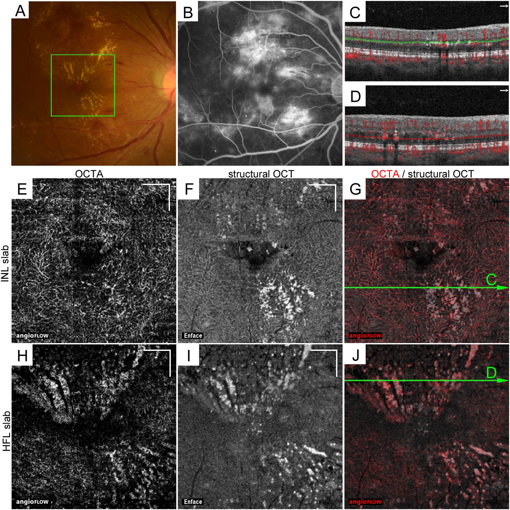
Decorrelation Signal of Diabetic Hyperreflective Foci on Optical Coherence  Tomography Angiography | Scientific Reports
Decorrelation Signal of Diabetic Hyperreflective Foci on Optical Coherence Tomography Angiography | Scientific Reports

Retinal Physician | PentaVision

Fig 3 | PLOS ONE
Fig 3 | PLOS ONE

Visualization with en face OCT offers value for ophthalmologists

Postoperative en face optical coherence tomography (OCT) scans of 12... |  Download Scientific Diagram
Postoperative en face optical coherence tomography (OCT) scans of 12... | Download Scientific Diagram
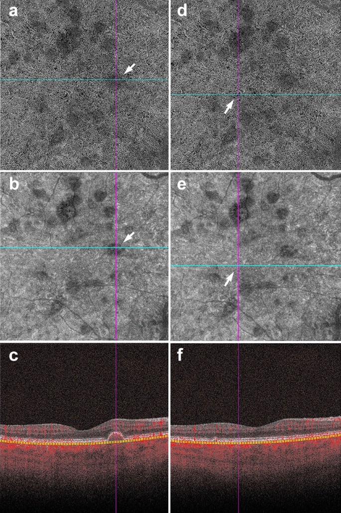
A practical guide to optical coherence tomography angiography  interpretation | International Journal of Retina and Vitreous | Full Text
A practical guide to optical coherence tomography angiography interpretation | International Journal of Retina and Vitreous | Full Text

PDF] En face optical coherence tomography of inner retinal defects after  internal limiting membrane peeling for idiopathic macular hole. | Semantic  Scholar
PDF] En face optical coherence tomography of inner retinal defects after internal limiting membrane peeling for idiopathic macular hole. | Semantic Scholar

En face OCT images generated by automated and manual segmentation at... |  Download Scientific Diagram
En face OCT images generated by automated and manual segmentation at... | Download Scientific Diagram

Evaluation of En-Face OCT Imaging in the Diagnosis of Macular Diseases

En face OCT images generated using the HD-OCT Zeiss Cirrus 5000. (A) En...  | Download Scientific Diagram
En face OCT images generated using the HD-OCT Zeiss Cirrus 5000. (A) En... | Download Scientific Diagram
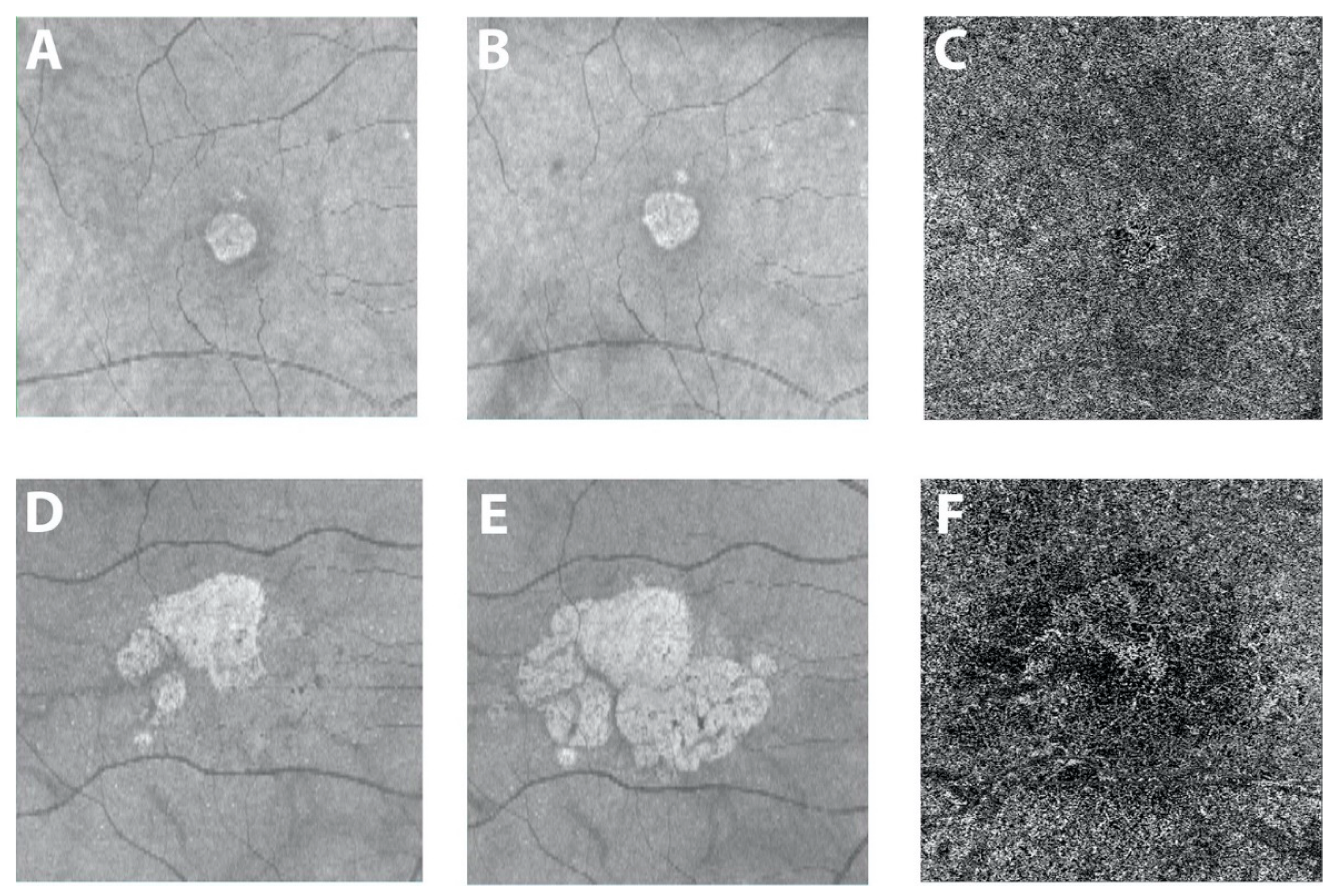
En Face OCT Superior to Red-free Imaging for RNFL Defects

JCM | Free Full-Text | Optical Coherence Tomography Angiography of the  Choriocapillaris in Age-Related Macular Degeneration
JCM | Free Full-Text | Optical Coherence Tomography Angiography of the Choriocapillaris in Age-Related Macular Degeneration

Comparison between OCTA and en-face OCT images in a healthy eye.: The... |  Download Scientific Diagram
Comparison between OCTA and en-face OCT images in a healthy eye.: The... | Download Scientific Diagram

En Face Optical Coherence Tomography Imaging of the Photoreceptor Layers in  Hydroxychloroquine Retinopathy - ScienceDirect
En Face Optical Coherence Tomography Imaging of the Photoreceptor Layers in Hydroxychloroquine Retinopathy - ScienceDirect

En Face Optical Coherence Tomography | Ento Key

Evaluation of retinal nerve fiber layer defect using wide-field en-face  swept-source OCT images by applying the inner limiting membrane flattening  | PLOS ONE
Evaluation of retinal nerve fiber layer defect using wide-field en-face swept-source OCT images by applying the inner limiting membrane flattening | PLOS ONE
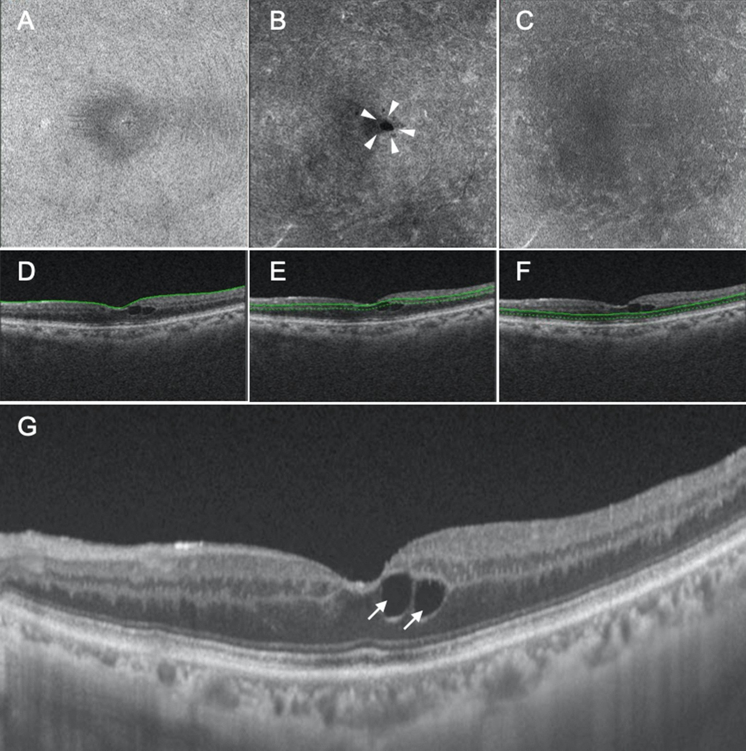
En-Face OCT Imaging | SpringerLink

En-Face OCT Imaging | SpringerLink

En face image-based classification of diabetic macular edema using swept  source optical coherence tomography | Scientific Reports
En face image-based classification of diabetic macular edema using swept source optical coherence tomography | Scientific Reports

Preoperative and postoperative features of macular holes on en face imaging  and optical coherence tomography angiography - ScienceDirect
Preoperative and postoperative features of macular holes on en face imaging and optical coherence tomography angiography - ScienceDirect

Use of En Face OCT to Identify Elevated ERM - Retina Today